
Clinical examination
This is one of the most important aspect of diagnosing ulcer infection. Clinical examination will help determine whether antibiotics are required and whether urgent surgical review should be sought. Ulcer infection produces cellulitis of the soft tissues, with erythema, warmth, oedema, purulent discharge, odour, increased exudate, and tenderness.
However, the classical features of infection may be diminished in patients with diabetes due to vasculopathy. In moderate infection lymphangitis may be present, in which linear erythematous streaking may be seen due to infection spreading via lymphatic vessels.
There may be changes in wound base characteristics from pink granulation tissue to a grey/yellow appearance with delayed or deteriorating healing despite adequate circulation and pressure off-loading. Inadequate assessment of clinical features at this point can lead to deeper and more invasive infection setting in.
The Infectious Disease Society of America (IDSA) has produced a grading system for clinicians, characterising the key features of infection, from mild to severe (table 2). This grading allows clinicians to decide on antibiotic type and formulation, and can also assist with decisions about hospital admission.
Table 2: Clinical characteristics of infection and severity based on definitions from the infectious Disease Society of America (2)
Uninfected
Fig: 1
No symptoms or signs of infection.
Mild Infection
Fig: 2
Local infection involving only the skin and subcutaneous tissue.
Erythema >0.5 cm to ≤2 cm around the ulcer
Excluding other causes of inflammatory response of the skin.
Moderate Infection
Fig: 3
Local infection with erythema >2cm, or involving structures deeper than the skin and subcutaneous tissues (e.g. abscess, osteomyelitis, septic arthritis, fascilitis) and with no systemic inflammatory response signs as in severe infection.
Severe Infection
Fig: 4
Local infection with the signs of systemic inflammatory response syndrome as characterised by ≥ 2 of the following:
Temp >38°C or < 36°C
Heart rate > 90 beats/min
Respiratory rate >20 breaths/min or PaCO2 <35mmHg
WBC >12000 or <4000 cells/mm3
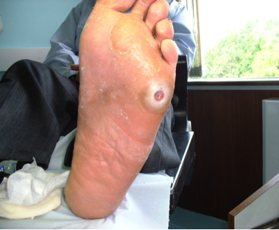
Figure 1. No Infection
Patient being managed for a wound on his foot with no clinical features of infection and systemically well. The wound does not probe to deeper sub-cutaneous layers or bone.
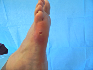
Figure 2. Mild infection
Patient attended with an infected foot wound which was showing deterioration with erythema around 2cms, not probing to bone but with increased exudate.
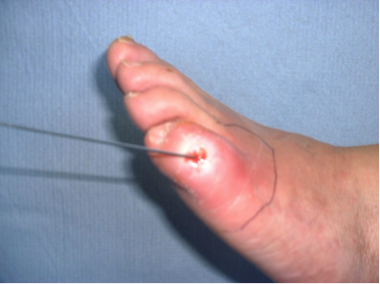
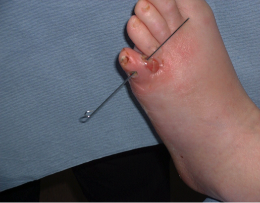
Figure 3. Moderate infection
This patient presented with a hot swollen toe which probed to bone, with erythema > 2cms.
The patient was well in himself but plain film x-rays confirmed the presence of osteomyelitis.
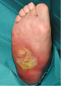
Figure 4. Severe infection
Patient presented with severe infected foot which was heavily exuding pus through the plantar aspect with a tracking bladder of pus to the lateral border of the foot. a foul smelling necrotic wound with moderate to heavy exudate.
On history taking they confirmed they were feeling feverish, nauseous and had noticed elevated blood sugar levels. Bloods revealed a CRP 242 mg/l.
Alternate layout
